A tooth can look healthy on the surface and still have decay developing where no patient can see it. That is what makes hidden cavities so easy to underestimate. Many people assume that if there is no obvious hole, no dark spot, and no pain, there is no real problem. Dentistry does not work that way. Cavities often begin between teeth, under older restorations, or inside grooves where early damage is easy to miss without the right evaluation. Dentists identify these hidden areas by combining visual judgment, imaging, surface assessment, and risk patterns rather than waiting for the damage to become obvious.
Why Some Cavities Stay Out Of Sight
- Where Early Decay Often Starts
Not all decay forms in places that are visible during normal brushing or a quick look in the mirror. Some cavities begin between teeth, where food debris and plaque remain trapped in narrow contact areas. Others develop beneath the surface of enamel that still appears mostly intact. That is why regular exams matter. A provider offering routine Dental Services is not only checking for visible damage. They are evaluating the areas where decay tends to progress quietly before symptoms appear.
- Visual Exams Still Matter First
Even though hidden cavities are not obvious to the patient, the dental exam still begins with direct observation. Dentists look for subtle changes in enamel color, texture, translucency, and contour. A tooth may not show a large visible cavity, but it may have chalky white areas, slight shadowing, or surface changes that suggest decay is developing beneath the surface. These small signs often guide the rest of the assessment.
This first step is important because experienced dentists are trained to notice patterns that patients would naturally miss. A tiny shift in how light reflects off enamel, or a slight discoloration around an old filling, can indicate that a closer look is needed. Hidden cavities are often first suspected through these quiet visual details before imaging confirms the full picture.
- X-Rays Reveal What Eyes Cannot.
One of the most useful tools for finding hidden cavities is dental radiography. X-rays allow dentists to see between teeth and beneath the visible enamel surface, where decay often develops long before it creates a visible opening. This is especially valuable for interproximal cavities, which form in the tight spaces where two teeth touch. These areas are among the most common sites for hidden decay because they are hard to inspect directly during a routine glance.
X-rays also help dentists evaluate whether decay is confined to the outer enamel or progressing deeper into the dentin. That distinction matters because the treatment approach varies with how far the damage has progressed. Without imaging, a dentist could miss early-stage decay that has not yet broken through to the visible outer surface.
- Surface Texture Offers Useful Clues
Dentists do not rely on color alone. They also evaluate how the tooth surface feels and behaves. Enamel affected by early decay may lose its normal hardness and become rougher or less stable in localized spots. Careful clinical assessment can help identify whether a groove, pit, or edge is holding plaque and beginning to weaken even before a cavity becomes obvious to the patient.
This part of the exam matters because hidden cavities do not always appear dramatically on imaging. In some cases, the combination of a suspicious texture, a plaque-retention pattern, and patient history gives the dentist sufficient reason to monitor an area more closely or recommend preventive steps before the damage progresses. Dentistry is often about catching small changes before they become structural problems.
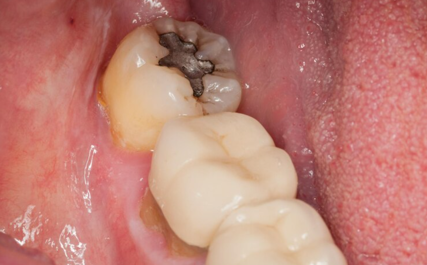
- Old Fillings Can Hide New Decay
Another common site for hidden cavities is around or beneath existing restorations. A tooth with an older filling may look stable from the outside, but decay can begin at the margins if bacteria enter through worn edges, tiny gaps, or material breakdown over time. Patients often assume that once a filling is placed, that tooth is no longer at risk. In reality, restored teeth still need regular evaluation because the surrounding structure can weaken without showing obvious external signs.
Dentists pay close attention to these areas during exams because recurrent decay often remains undetected until it becomes more extensive. Imaging, margin inspection, and careful clinical judgment all help determine whether the restoration is still sealing the tooth well or whether hidden damage may be forming underneath.
Why Early Detection Makes Treatment Easier
Dentists identify hidden cavities by looking beyond what is obvious on the tooth surface. They use visual examination, X-rays, surface evaluation, restoration checks, patient risk patterns, and supporting diagnostic tools to find decay before it becomes large enough to cause pain or visible damage. That approach matters because cavities are much easier to manage when they are found early.
For patients, the practical lesson is simple. A tooth does not need to look damaged for decay to start. Regular exams give dentists the chance to detect those hidden areas while treatment is still more conservative and the tooth structure is better preserved.